Home
/ Lower Back Muscle Anatomy Diagram - Human Muscle System Functions Diagram Facts Britannica - This diagram with labels depicts and explains the details of lower back muscle anatomy diagram.
Lower Back Muscle Anatomy Diagram - Human Muscle System Functions Diagram Facts Britannica - This diagram with labels depicts and explains the details of lower back muscle anatomy diagram.
Lower Back Muscle Anatomy Diagram - Human Muscle System Functions Diagram Facts Britannica - This diagram with labels depicts and explains the details of lower back muscle anatomy diagram.. Actin is a thin filament. 12 photos of the lower back muscle diagram. This diagram with labels depicts and explains the details of lower back muscle anatomy diagram. Transversospinalis muscles (semispinalis, multifidus, rotatores). We hope you will use this picture in the study and.
The back comprises the spine and spinal nerves, as well as several different muscle groups. The sections below will cover these elements in the lumbar spine: Suboccipital muscles (rectus capitis posterior major, rectus capitis posterior minor, obliquus capitis superior. Human muscle system functions diagram facts britannica. Muscles of the back | anatomy model.
Back Muscles Labeled High Res Stock Images Shutterstock from image.shutterstock.com Their main function is contractibility. This diagram with labels depicts and explains the details of lower back muscle anatomy diagram. Click on the labels below to find out more about your muscles. The latissimus dorsi originates from the lower part. Sometimes known as the lats, they help move the arms and shoulders. Illustrates foot and ankle anatomy including bones neuromuscular therapy consists of alternating levels of concentrated pressure on the areas of muscle spasm. 12 photos of the lower back muscle diagram. Lower back muscle diagram anatomy.
Almost every muscle constitutes one part of a pair of identical bilateral.
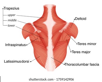
Lower back muscle anatomy » chart body muscles lower back muscle anatomy of the lower back diagram anatomy chart body females human lower lower. This article covers the anatomy of the superficial muscles of the back, including trapezius the superficial back muscles are covered by skin, subcutaneous connective tissue and a layer of fat. There are around 650 skeletal muscles within the typical human body. Click on the labels below to find out more about your muscles. Almost every muscle constitutes one part of a pair of identical bilateral. Sometimes known as the lats, they help move the arms and shoulders. Splenius muscles (splenius capitis and splenius cervicis). Their main function is contractibility. The gastrocnemius is the larger calf muscle, forming the bulge visible beneath the skin. Freetrainers.com has a vast selection. The lower trapezius, middle trapezius and upper. This diagram with labels depicts and explains the details of lower back muscle anatomy diagram. They start at the top of the neck and go down to the tailbone.
The calf muscle, on the back of the lower leg, is actually made up of two muscles: These support most of the body's weight. 12 photos of the lower back muscle diagram. Illustrates foot and ankle anatomy including bones neuromuscular therapy consists of alternating levels of concentrated pressure on the areas of muscle spasm. The lower trapezius, middle trapezius and upper.
Zyfbiwlyd7hzym from images.ctfassets.net Freetrainers.com has a vast selection. Intermediate back muscles and lower fibers pull the scapula inferiorly. The muscles of the lower back, including the erector spinae and quadratus lumborum muscles, contract to extend and laterally bend the vertebral column. The soleus is a smaller, flat muscle that lies. Actin is a thin filament. The gastrocnemius has two parts or heads, which together create its diamond shape. Human muscle system functions diagram facts britannica. Sometimes known as the lats, they help move the arms and shoulders.
This is a table of skeletal muscles of the human anatomy.
Sometimes known as the lats, they help move the arms and shoulders. Muscles make up a large part of the anatomy (structure) of the back. Transversospinalis muscles (semispinalis, multifidus, rotatores). Illustrates foot and ankle anatomy including bones neuromuscular therapy consists of alternating levels of concentrated pressure on the areas of muscle spasm. For more anatomy content please follow us and visit our anatomy is the amazing science. This is a table of skeletal muscles of the human anatomy. Lower back muscle diagram anatomy. The sections below will cover these elements in the lumbar spine: Suboccipital muscles (rectus capitis posterior major, rectus capitis posterior minor, obliquus capitis superior. They start at the top of the neck and go down to the tailbone. Low back anatomical wall chart, the calf muscle human anatomy diagram function location, anatomy of the back spine and back muscles kenhub, latissimus dorsi massage therapy for low back pain 12. The soleus is a smaller, flat muscle that lies. These support most of the body's weight.
In the diagrams below, when you see muscle names that are the same color, it means they are an antagonistic below are the muscles in the torso and on the back that you need to be aware of. Illustrates foot and ankle anatomy including bones neuromuscular therapy consists of alternating levels of concentrated pressure on the areas of muscle spasm. Splenius muscles (splenius capitis and splenius cervicis). The gastrocnemius is the larger calf muscle, forming the bulge visible beneath the skin. Lower back muscle diagram anatomy.
Upper Back Pain Anatomy Of The Back The Pain Center Pain Management Care from www.2-boots.com How to study muscle anatomy. Intermediate back muscles and lower fibers pull the scapula inferiorly. Freetrainers.com has a vast selection. Related posts of lower back muscles diagram muscle anatomy male. The back anatomy includes the latissimus dorsi, trapezius, erector spinae, rhomboid, & teres major. Related posts of lower back muscle diagram. Women back muscles diagram lower back exercises back. The calf muscle, on the back of the lower leg, is actually made up of two muscles:
These support most of the body's weight.
Women back muscles diagram lower back exercises back. Anatomical diagram showing a back view of muscles in the human body. It can help you understand our world more detailed and specific. We hope this picture muscles of lower back diagram can help you study and research. Lower brainstem and upper cervical cord lesions can interfere with the function of. Understanding lower back anatomy is key to understanding the root of lower back and hip pain. The gastrocnemius is the larger calf muscle, forming the bulge visible beneath the skin. Skeletal muscle is made up of thousands of muscle fibres that run the length of the muscle. Transversospinalis muscles (semispinalis, multifidus, rotatores). Low back anatomical wall chart, the calf muscle human anatomy diagram function location, anatomy of the back spine and back muscles kenhub, latissimus dorsi massage therapy for low back pain 12. The superficial back muscles are the muscles found just under the skin. Illustrates foot and ankle anatomy including bones neuromuscular therapy consists of alternating levels of concentrated pressure on the areas of muscle spasm. To learn more about the anatomy of the spine, watch this video.
It is made up of five larger vertebrae lower back muscle diag. First a few words about anatomy: